08 Aug

Eye Donation: What you need to know and do – Part 2
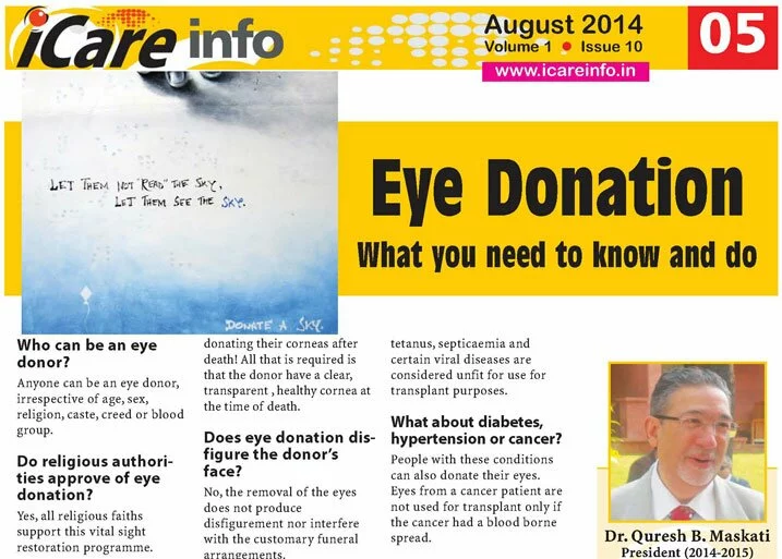
i-Care-Info-Eye-Donation-What-you-need-to-know-and-do-Maskati.jpg

What is to be done when a relative expires?

  1. Keep the death certificate or its copy to show to the eye bank team. They do not need to take it away. You do not need to look for a photocopier machine to make a photocopy in the middle of the night. The eye bank team only needs to look at the certificate to determine:
    •  The patient is really dead and
    •  The cause of death, to determine if the eyeball can be useful.
  2. Contact your nearest eye bank – in India, most cities have 1919 as the eye bank contact number. Otherwise, check the local telephone directory – most eye banks are listed in the emergency numbers list. The eye bank team will arrive in the next 30-60 minutes, provided you have given them an accurate address with landmarks. The procedure will take only around 30-40 minutes.
  3. Close the eye lids, while waiting for the eye bank team and put off any overhead fan, to prevent drying of the cornea. However, if there is an air-conditioner, switch it on, if possible.
  4. Cover the closed lids with moist cotton wool (if readily available in the house, otherwise do not bother).
  5. Raise the donor’s head by 6 inches, by placing 2 pillows under it.

Is there any cumbersome paperwork or time consuming formalities?
None whatsoever. The eye bank team will come with a form, which they will fill themselves. They will only need the signatures of 2 close relatives, giving consent. It does not matter whether the donor had pledged his eyes for donation during his lifetime or not.

I believe some blood is also collected?
Yes, according to WHO guidelines, the team will also collect some blood from the donor, for testing for HIV, hepatitis etc. They will withdraw this from a large vessel or from the heart with a needle puncture.

Is there a real need for Eye Donation?
Yes, in India there are lacs of patients awaiting corneal transplant.. in some eye banks patients wait over a year for their turn. Every year barely 30,000 corneas are donated, so the waiting list keeps growing!

Finally, is there any money to be paid?
No, no monetary transaction is done. Most eye banks recover their running costs by charging a small processing fee to the recipients.

If it does not cost to donate eyeballs after death and the procedure is so easy, and encouraged by every religion, why are there such few donors?
Good question. You tell me! You go around spreading the message of the nobility of eye donation and how easy and hassle free it is and maybe by the time I write next year, this question will need deletion!

 Eye Donation: What you need to know and do – Part 1


08 Aug

Eye Donation: What you need to know and do – Part 1
i-Care-Info-Eye-Donation-What-you-need-to-know-and-do-Maskati.jpg

Who can be an eye donor?
Anyone can be an eye donor, irrespective of age, sex, religion, caste, creed or blood group.

Do religious authorities approve of eye donation?
Yes, all religious faiths support this vital sight restoration programme.

Is the whole eye used for transplant?
No, only the thin transparent layer in front of the iris, called the cornea is used for transplant, to replace the diseased or opaque cornea in the eye of the patient (recipient).

Do cataracts or the use of spectacles render the cornea unfit for donation?
No, both these conditions relate to the lens of the eye and not the cornea. In fact, people who have been successfully operated for cataracts or glaucoma or even retinal detachments can also donate their eyes after death. In fact there have been instances of those who have received a corneal transplant themselves donating their corneas after death! All that is required is that the donor have a clear, transparent , healthy cornea at the time of death.

Does eye donation disfigure the donor’s face?
No, the removal of the eyes does not produce disfigurement nor interfere with the customary funeral arrangements.

Does the human body reject the transplanted donor’s cornea? How successful are corneal transplants?
The human cornea does not have any blood supply; hence the risk of rejection is very low. Rejection, if it does occur can be suppressed by timely medication. In general, the chances of success are greater than 80%. In deep anterior lamellar transplants, where the inner lining of the cornea called the endothelium, is not transplanted, the success rate is as high as 95%!

What conditions render the cornea unfit for donation?
Corneas of patients suffering from AIDS, rabies, syphilis, tetanus, septicaemia and certain viral diseases are considered unfit for use for transplant purposes.

What about diabetes, hypertension or cancer?
People with these conditions can also donate their eyes. Eyes from a cancer patient are not used for transplant only if the cancer had a blood borne spread.

Is there any use for corneas which are for some reason unfit for transplant?
Corneas, which for some reason cannot be used for vision restoring corneal transplants, are invaluable for research and training programmes.

How will my donation be used?
After the eyes are removed, they will be evaluated, processed, screened and then supplied to the eye surgeon for transplant.

Do corneal transplants guarantee sight to all blind people?
No, transplants only help when the loss of sight is solely due to corneal defects and the rest of the eye mechanism is intact.

How quickly should eyes be removed after death?
As soon as possible, but eyes can be removed up to 6 hours after death. However, in places where the climate is hot such as most parts of India, a shorter duration, preferably 2 to 4 hours after death is advisable.

Is it necessary to transport the donor to the hospital after death?
No, eye banks have personnel who will come to the donor’s home or place of death and remove the eyes.

What is an eye bank?
An eye bank is the link between the donor and recipient/eye surgeon. It is an organization recognised by the government to collect and distribute human eyes to those who require corneal transplants.

Eye Donation: What you need to know and do – Part 2

  • What is to be done when a relative expires?
  • Is there any cumbersome paperwork or time consuming formalities?
  • I believe some blood is also collected?Is there a real need for Eye Donation?
  • Finally, is there any money to be paid?
  • If it does not cost to donate eyeballs after death and the procedure is so easy, and encouraged by every religion, why are there such few donors?


10 Jul

Common Investigations of Your Eye – Part 1

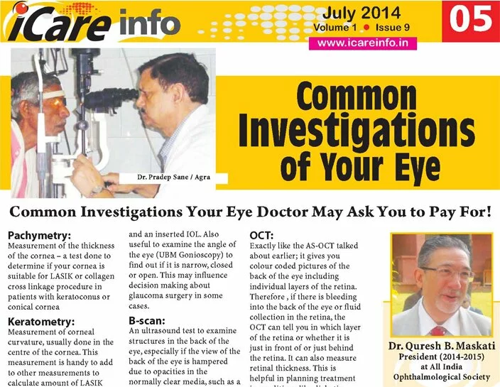
iCare-Info-Eye-Test-Maskati

Common Investigations Your Eye Doctor May Ask You to Pay For!

Pachymetry:
Measurement of the thickness of the cornea – a test done to determine if your cornea is suitable for LASIK or collagen cross linkage procedure in patients with keratoconus or conical cornea

Keratometry:
Measurement of corneal curvature, usually done in the centre of the cornea. This measurement is handy to add to other measurements to calculate amount of LASIK laser required to bring your spectacle number to zero; also one of the measurements required to calculate the Intra-ocular lens power suitable for your eye in case you have a cataract.

AS-OCT:
Anterior Segment Optical Coherence Tomography. Something like a CT scan of the front portion of the eye. Comes in handy for a variety of diseases of the cornea and anterior segment

UBM – Ultrabiomicroscopy:
This is an ophthalmic ultrasound using a special probe, to view in detail structures in the anterior segment, up to and including the crystalline lens of the eye and an inserted IOL. Also useful to examine the angle of the eye (UBM Gonioscopy) to find out if it is narrow, closed or open. This may influence decision making about glaucoma surgery in some cases.

B-scan:
An ultrasound test to examine structures in the back of the eye, especially if the view of the back of the eye is hampered due to opacities in the normally clear media, such as a cataract or haemorrhage in the posterior compartment (vitreous haemorrhage), so that ophthalmoscopy examination is not possible. Also useful in lesions of the extra-ocular muscles and any space occupying lesions in the orbit, outside the eye.

A-scan:
Also called A scan biometry. In this the examiner determines the front to back measurement of the eye using ultrasound. He then feeds in the keratometry reading obtained from the keratometer. An inbuilt computer in the A- scan biometry machine, using one of several complex formulae, then calculate the power of the intra-ocular lens implant required for your eye

OCT:
Exactly like the AS-OCT talked about earlier; it gives you colour coded pictures of the back of the eye including individual layers of the retina. Therefore , if there is bleeding into the back of the eye or fluid collection in the retina, the OCT can tell you in which layer of the retina or whether it is just in front of or just behind the retina. It can also measure retinal thickness. This is helpful in planning treatment in conditions like diabetic macular oedema or monitoring efficacy of intravitreal injections given for wet age related macular degeneration.

Corneal Topography:
This gives you several ‘maps’ of the cornea; an invaluable tool in assessing suitability of the cornea for LASIK and useful in detection of keratoconus and in fitting of contact lenses. It provides information on the curvature of the cornea at various points of the cornea, the ‘power’ of the cornea etc.

Aberrometry:
This is often used by the LASIK surgeon in patients unhappy after a successful LASIK – it detects minute flaws called “higher order aberrations” in your eye’s optical system which prevents you from enjoying ‘super’ vision after LASIK. Some LASIK machines which are “wave-front guided” routinely use data from aberrometers to fine tune the LASIK treatment to reduce these aberrations post LASIK surgery.

Visual Field Analysis:
Mechanical devices that measured your field of vision were called perimeters. They have been largely replaced by Visual Field Analysers (VFA). These have inbuilt computers for storing and analysing your visual fields. They can compare fields done over a period and analyse whether the patient’s field is worsening or remaining the same. The routine test is a little cumbersome and requires your co-operation, lasting around 30-40 minutes, though for simple screening faster tests are available in the same machine. VFA is ordered in cases of glaucoma both for diagnosis and for judging efficacy of treatment. They are also invaluable in some neurological disorders causing drop in vision like in stroke etc.

Common Investigations  of Your Eye – Part 2


10 Jul

Common Investigations of Your Eye – Part 2

iCare-Info-Eye-Test-Maskati

Common Investigations Your Eye Doctor May Ask You to Pay For!

Fluorescein Fundus Angiography (FFA):
In this test a dye, fluorescein Sodium is injected into the vein of your forearm. It takes around 10 seconds for the dye to spread into your retinal vasculature. This can be viewed by the observer using an ophthalmoscope, though more commonly, a digital camera called a fundus camera is used to take multiple photos of the retina shortly after dye injection.
Depending on the areas of perfusion (dye seen) or non-perfusion (dye not seen) or leakage of dye from the vessels into the retina, decisions are made in cases of diabetic retinopathy, age-related macular degeneration etc. about whether to do laser or give some injections or treat with medications .

HRT/GDX :
These are fancy names for computer software in which images of the optic disc and retina are analysed. Accurate measurements are made of optic nerve size, cup/disc ratio, retinal nerve fibre thickness. This is a useful adjunct to diagnose glaucoma and monitor its progression.

Electro-retinography (ERG):
This test is used to find out the function of the retina. This comes in useful when the retina appears normal on ophthalmoscopy but patient has poor vision and also when the retina is not clearly seen due to media opacities. If the ERG is poor or absent, the eye surgeon may decide that the visual prognosis is poor and further surgery to clear the media opacities like a corneal transplant for corneal opacities or a cataract extraction for a mature cataract is not warranted.

M-ERG or Multifocal ERG:
This is the newest version of the ERG. In this a colour map is generated with the area of maximum cone function (macula) showing a steep elevation while the area with no cones (optic nerve ) showing up as flat and blue in colour. In diseases like macular degeneration, the central hill is considerably flattened, making diagnosis a ‘no-brainer’.

Visual Evoked Potential (VEP):
In this test, either a very bright light (Flash VEP) or a checker board pattern is shone into the patient’s eye. The time taken for this to be perceived by the visual area of the brain (situated at the back of the head) and the intensity of the signal is recorded by sensitive electrodes placed at the back of the scalp. This measures the integrity of the visual pathway from the eye right up to the brain. In conditions like optic neuritis, where the optic nerve which carries signals from the eye to the brain is inflamed, the VEP will be reduced. On the other hand, a patient who is feigning blindness will have a normal VEP.

Can a person who has undergone one or more of these tests donate their eyes?

None of these tests is life threatening. However, it is proved that every patient who undergoes any of these tests will die some day, maybe months, years or decades later! If a patient were to die due to natural or accidental causes years after any of these tests were performed, the author does recommend donation of the eyes!

Common Investigations  of Your Eye – Part 1